How It Works
A simple, four-step process to enhance your clinical workflow
Capture & Upload
- Fundus Image
- OCT Scan
- External Eye Photo(compatible with any digital camera including mobile)
AI Analysis
Our AI models act as a second set of eyes, supporting your expertise by highlighting potential areas for investigation while leaving the final assessment in your hands.
Review
Optometrists and eye-care professionals can review AI findings, add their own insights, and share results with patients or fellow specialists.
Improve
Continuous updates to our AI ensure improved accuracy and new capabilities. New algorithms added regularly.
Seamless Integration into Your Practice
Our AI assistant works alongside your existing workflow, enhancing your clinical capabilities without disrupting your established processes. You maintain full control while gaining valuable insights.
Continue your normal appointment booking and patient intake process. No changes needed to your existing scheduling system or initial patient interactions.
During your standard examination, simply capture images as you normally would. Our software works with your existing imaging formats, requiring no additional hardware investment.
While you conduct your professional assessment, our AI quickly analyzes the images in the background, preparing additional insights for your review. You maintain complete control over the clinical process.
Review the AI findings alongside your professional assessment. Use the additional insights to support your clinical evaluation. The final decision always remains in your expert hands.
Use our intuitive pdf document to explain findings to patients, showing them exactly what you have identified. This helps improve patient understanding with treatment plans.
Key Benefits
Transform your practice with AI-powered eye care technology
Early Analysis & Screening
Swiftly identify potential findings related to common eye conditions, highlighting areas of interest for clinician review.
Revenue Growth
Increase practice revenue through premium AI-enhanced screenings with flexible pay-per-use pricing.
Evidence-Based Insights
Provide a second opinion backed by deep-learning algorithms, giving greater confidence in clinical decision-making.
Seamless Integration
Compatible with existing imaging devices and clinic workflows; minimal disruption, maximum value.
See AI Analysis in Action
See how our AI technology identifies and analyzes various eye conditions from standard images.
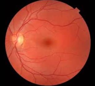
Fundus Image Analysis
Key Observations
- Retina: Healthy appearance with well-defined layers
- Optic Nerve: Intact with normal appearance
- Blood Vessels: Normal with no visible abnormalities
- Macula: Normal central vision area
Assessment
No signs of diabetic retinopathy, retinal detachment, or age-related macular degeneration.
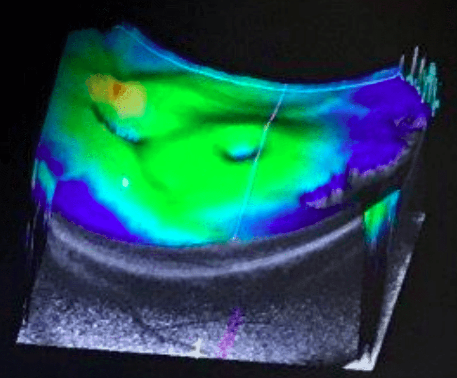
OCT Scan Analysis
Key Observations
- Cross-section: Clear visualization of retinal layers
- Retinal Thickness: Normal and consistent
- Optic Nerve: No compression or damage
- Macular Region: Well-defined with no abnormalities
Assessment
Healthy eye structure with no indications of macular degeneration, edema, or retinal disorders.
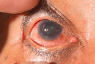
External Eye Analysis
Highlighted Findings
- Anterior Chamber: Fluid accumulation present
- Retina: Irregular with scattered hemorrhages
- Blood Vessels: Showing tortuosity and damage
- Macula: Signs of edema and irregularity
Areas of Interest
Potential findings requiring further clinical evaluation by eye care professional.
Targeted at
Empowering eye care professionals across all settings
Optometrists & Ophthalmologists
Gain a powerful AI assistant to help with everyday screenings, store patient records securely, and quickly spot irregularities.
Hospitals
Streamline patient flow and reduce wait times by triaging complex cases faster. Integrates easily into hospital-wide health record systems.
Clinics & Telemedicine
Expand remote screening capabilities and maintain high standards no matter where your patients are.
Important Notice
This tool provides AI-assisted analysis for clinician review. It is not intended for diagnostic or treatment decisions.
AI-Assisted Eye Image Analysis
Join leading eye care professionals who trust our AI-assisted decision support tool to highlight potential findings for clinician review.
This tool provides AI-assisted analysis for clinician review. It is not intended for diagnostic or treatment decisions.